
A cancer diagnosis rarely rests on one doctor's opinion. Behind most treatment decisions sits a multidisciplinary team, a group of specialists who each bring a different lens to the same case. Imaging is one of the few pieces of information every specialist at that table relies on, from the oncologist planning treatment to the surgeon deciding what's operable. Without accurate, well-timed imaging, that team can't do its job properly.
This kind of coordinated, team-based approach has become the standard model for cancer care across Australia, replacing the older pattern of a patient moving between specialists one at a time. Understanding how imaging fits into that process helps explain why scan timing, quality, and reporting speed matter so much to your overall care outcome.
What Is a Multidisciplinary Cancer Care Team?
A multidisciplinary team, often shortened to MDT, is a group of healthcare professionals from different specialties who meet regularly to discuss a patient's cancer diagnosis and treatment plan together. A typical MDT includes a medical oncologist, surgeon, radiation oncologist, pathologist, radiologist, and often a specialist nurse or care coordinator.
Instead of a patient seeing each specialist separately and hoping their advice aligns, the team reviews the same case file together. This reduces the chance of conflicting recommendations and speeds up the path from diagnosis to treatment.
Why Imaging Is Central to MDT Decisions
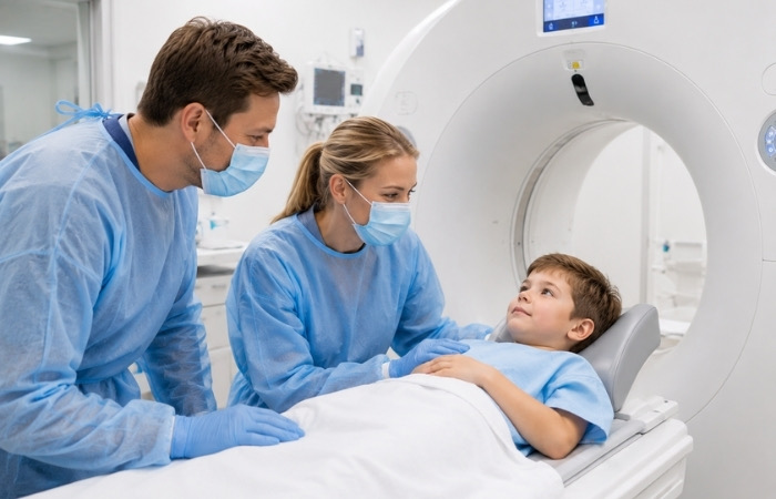
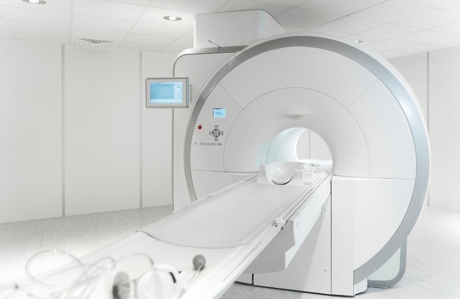
Each scan type works differently and is suited to different symptoms, from soft tissue concerns to suspected fractures.
Why Imaging Is Central to MDT Decisions
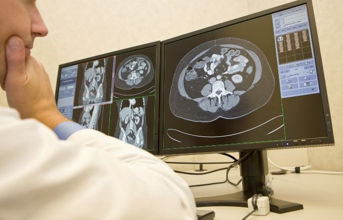
Imaging gives the MDT a shared, objective starting point. A scan doesn't carry the bias of a single specialist's perspective. It shows tumour size, location, and whether cancer has spread, information that shapes almost every decision made in the room.
For a surgeon, imaging determines whether a tumour is operable and what approach makes sense. For a radiation oncologist, it defines the treatment field. For a medical oncologist, it establishes a baseline to measure whether treatment is working. Pathology tells the team what the cancer is at a cellular level, while imaging tells them where it is and how far it has progressed. Both pieces of information are needed to build a coordinated plan.
This is also why timing matters so much. An MDT meeting works best when every specialist has current imaging in front of them. Outdated or incomplete scans can delay a case being discussed or force a decision to be made with an incomplete picture.

The Radiologist's Specific Contribution
While every specialist at the table brings a distinct skill set, the radiologist's role is often less visible to patients than the surgeon's or oncologist's, even though it sits underneath almost every other decision made in the room.
A radiologist doesn't just take a scan and hand over an image. They interpret what that image shows, measure tumour dimensions precisely, compare current scans against previous ones to track change over time, and flag anything unexpected that might change the direction of the discussion. In many MDT meetings, the radiologist presents the imaging findings directly to the rest of the team, walking through what the scan shows and answering questions in real time.
This interpretive layer is what turns a raw scan into something the rest of the team can actually act on. Two people can look at the same image, but only a trained radiologist can translate it into the specific measurements and observations an oncologist or surgeon needs to plan treatment.
How Imaging Feeds Into the Decision-Making Process

- Initial diagnostic imaging. A scan such as a CT, ultrasound, or X-ray is typically what identifies a suspicious finding in the first place, prompting referral for further investigation.
- Staging. Once cancer is suspected or confirmed, further imaging helps determine how advanced it is, including whether it has spread to nearby tissue, lymph nodes, or other organs.
- MDT review. The imaging, alongside pathology results and the patient's clinical history, is presented to the full team, who discuss treatment options together.
- Treatment planning. Surgeons, radiation oncologists, and medical oncologists use the imaging to map out their specific approach, whether that's surgical planning, radiation field design, or systemic therapy.
- Image-guided procedures. In many cases, a biopsy or other tissue sample is taken using imaging guidance, ensuring the right area is targeted with precision.
- Monitoring. Follow-up imaging tracks how a tumour responds to treatment, giving the team objective evidence to continue, adjust, or conclude a course of treatment.
Each of these steps depends on accurate, well-reported imaging. A missed detail at the diagnostic stage can carry through the entire treatment pathway.
Imaging Modalities Commonly Used in Cancer Care
Different imaging tools serve different purposes across the cancer care journey. Here's a general comparison of what each is typically used for.
| Modality | Primary Role in Cancer Care | Typical Use Case |
|---|---|---|
| CT scan | Detailed cross-sectional imaging | Staging, treatment planning, monitoring |
| Ultrasound | Real-time, radiation-free imaging | Guiding biopsies, assessing soft tissue |
| X-ray | Basic structural imaging | Initial checks, some lung findings |
| Low-dose CT | Lung-specific screening | Early detection in high-risk patients |
| Image-guided biopsy | Tissue sampling with imaging guidance | Confirming diagnosis, cellular analysis |
The right modality depends on the type of cancer being investigated, the stage of care, and what specific question the MDT needs answered. Your referring doctor and the imaging team work together to determine which scan is appropriate at each stage.
A Practical Example of Imaging in Lung Cancer Pathways
Lung cancer offers a clear example of how this process plays out. A patient considered high risk, often due to smoking history or family history, may undergo a low-dose CT lung screening scan. If that scan identifies a suspicious nodule, the next steps typically involve further imaging to characterise it, followed by a biopsy if needed to confirm what the tissue actually is.
From there, the case moves to an MDT, where the radiologist presents the imaging findings alongside the pathology results. The surgeon assesses whether the nodule or tumour is operable based on its location and size. The radiation oncologist considers whether radiotherapy is appropriate, and the medical oncologist weighs up systemic treatment options. Every one of these conversations references the same set of images, which is why consistency and accuracy at the imaging stage carry so much weight for what happens next.

This pathway also illustrates why using the same imaging provider across a patient's care journey has practical value. Comparable scans, taken on consistent equipment and reported with a consistent frame of reference, make it easier for the MDT to track subtle changes over time rather than relying on scans that vary in quality or format.
Where Diagnostic Imaging Clinics Fit Into the Bigger Picture
A diagnostic imaging clinic is often the first point of contact in this entire process, well before a patient ever sits in front of an MDT. It's where the initial images that flag a concern and start the referral pathway actually get produced, and where any tissue sampling needed to confirm a diagnosis typically takes place.
Fast, accurate reporting at this stage matters enormously. An MDT can only meet as often as the imaging and pathology results allow, so a delay in reporting can push back the entire treatment timeline. Clear communication between the imaging clinic, the referring doctor, and the broader care team helps keep that pathway moving.
Frequently Asked Questions
What does MDT stand for in cancer care?
MDT stands for multidisciplinary team, a group of specialists from different fields who meet to discuss and plan a patient's cancer care together, rather than working in isolation.
How often does an MDT review a patient's imaging?
This varies depending on the type of cancer and stage of treatment. Some patients are discussed at diagnosis and again at key treatment milestones, while others may be reviewed more frequently if their case is complex or their treatment response needs closer tracking.
Can a patient attend their own MDT meeting?
In most cases, MDT meetings are a clinical discussion among specialists rather than a patient-facing appointment. Your treating doctor will usually explain the outcome and next steps to you directly afterwards, translating the clinical discussion into a plan you can act on.
Why is imaging repeated during treatment?
Repeat imaging gives the MDT objective evidence of how a tumour is responding, which helps determine whether the current treatment plan should continue, change, or conclude. It also helps confirm whether earlier findings were accurately assessed.
Does every cancer diagnosis go through an MDT?
Most complex or newly diagnosed cancers are discussed at an MDT meeting, though the exact process can vary between hospitals and cancer types. Your specialist can explain how your particular case is being managed.
Supporting Better-Informed Cancer Care Decisions
Imaging doesn't sit on the sidelines of cancer care; it's one of the pillars the entire multidisciplinary process is built on. From the first scan that flags a concern through to the follow-up imaging that tracks treatment response, accurate and timely imaging keeps the whole team working from the same picture.

This matters just as much for the person behind the scan as it does for the specialists reading it. A cancer diagnosis already brings enough uncertainty, and knowing that your care is being shaped by a coordinated team working from consistent, clearly reported imaging can make that process feel less fragmented. It also means fewer repeat scans, faster movement from one stage of care to the next, and a treatment plan that reflects genuine agreement across specialties rather than a single opinion. For referring doctors, it means confidence that the imaging they're building a case on will hold up to scrutiny from every other specialist in the room.
Contact us to book a referral-based scan and support your care team with clear, timely imaging.