Acute abdominal pain arrives without warning. It can signal anything from a kidney stone to appendicitis, bowel obstruction, or a ruptured ovarian cyst. The range of possible causes is broad, the overlap in symptoms is real, and getting the right imaging at the right time makes a meaningful difference to how quickly and accurately a diagnosis is reached.
Not every scan suits every cause. An ultrasound won't reliably show a perforated bowel. An X-ray won't reveal an inflamed gallbladder. The scan that gets ordered matters, and understanding why one is chosen over another helps you make sense of what your doctor is recommending.
What Counts as Acute Abdominal Pain
Acute abdominal pain is pain in the abdomen that comes on suddenly and is severe enough to require medical assessment. It covers a wide range of presentations: pain that is sharp, cramping, constant, or radiating; pain that begins in one location and spreads; or pain that is diffuse from the start.
Common causes include:
- Appendicitis
- Gallstones or cholecystitis (inflammation of the gallbladder)
- Kidney stones (renal colic)
- Bowel obstruction
- Diverticulitis
- Pancreatitis
- Perforated peptic ulcer
- Ovarian cysts or ectopic pregnancy
- Hernia complications
The location, character, and onset of pain all give clinicians useful information, but symptoms frequently overlap. Imaging is often needed to narrow the diagnostic possibilities and guide treatment decisions.
Why the Right Scan Matters
Ordering the wrong scan does not just delay a diagnosis. It can mean repeat imaging, additional radiation exposure, or a missed finding that changes the clinical picture. Each imaging modality has clear strengths and recognised limitations, and choosing between them depends on what is most likely causing the pain, where in the abdomen it originates, and what clinical information is already available.
Speed matters too. In acute presentations, the time between symptom onset and diagnosis affects outcomes for conditions like appendicitis, bowel ischaemia, and ectopic pregnancy. Imaging that delivers rapid, high-quality information about the likely cause shortens that diagnostic window meaningfully.
The Three Main Scans Used for Acute Abdominal Pain
CT Scan

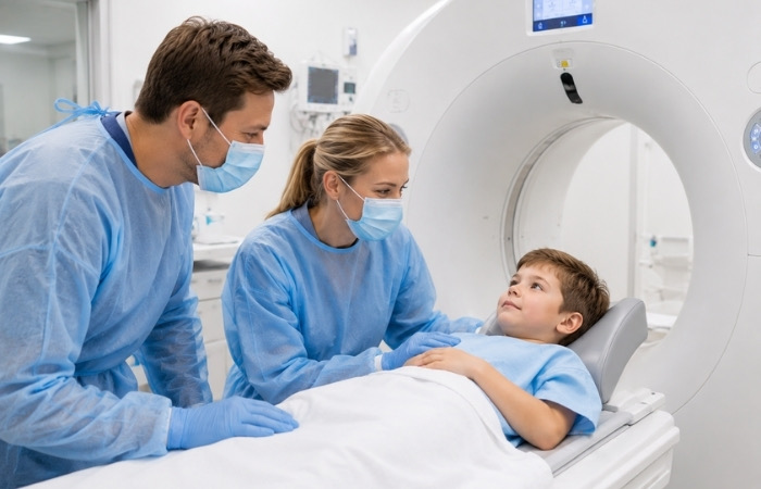
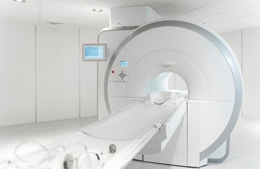
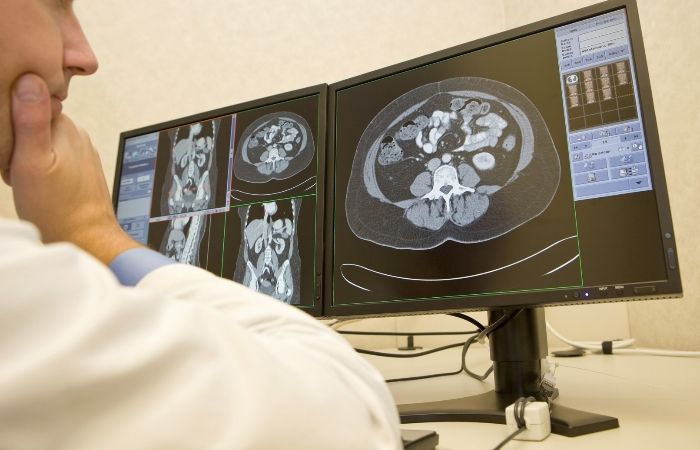
A CT (computed tomography) scan uses X-ray beams from multiple angles to produce detailed cross-sectional images of the abdomen and pelvis. It is the most comprehensive first-line imaging tool for acute abdominal pain in adults when the cause is unclear, providing information about solid organs, bowel, blood vessels, lymph nodes, and surrounding structures in a single examination.
CT is particularly well-suited to suspected appendicitis, diverticulitis, bowel obstruction, bowel perforation, kidney stones, and abdominal aortic aneurysm. It can be performed with or without intravenous contrast depending on what is being investigated.
CT does involve radiation exposure, which is a factor weighed by the referring doctor, particularly in younger patients. The clinical benefit of accurate, timely diagnosis is considered alongside this factor on an individual basis.
Ultrasound

Ultrasound uses high-frequency sound waves to produce real-time images of internal structures. It does not use radiation, making it the preferred first-line investigation for suspected gallbladder disease, kidney conditions, and abdominal pain in pregnant women.
Ultrasound is well-suited to assessing the gallbladder for stones and inflammation, the kidneys and urinary tract, the liver and spleen, and pelvic structures including the ovaries and uterus. It is widely available and can often be performed quickly.
Its limitations are worth understanding. Image quality can be affected by body habitus, bowel gas, and patient positioning. Ultrasound is less reliable for visualising the bowel itself or retroperitoneal structures, which is why CT is often preferred when a broader assessment is needed.
X-Ray

An abdominal X-ray is a fast, low-cost imaging tool with a specific but limited role in acute abdominal pain. It is most useful for identifying bowel obstruction (shown by dilated loops of bowel), bowel perforation (shown by free air under the diaphragm on an upright chest X-ray), and certain radiopaque foreign bodies or kidney stones.
X-ray provides limited information about soft tissue structures and cannot reliably visualise the appendix, gallbladder, or most causes of pelvic pain. In many acute presentations, it has been largely superseded by CT for diagnostic purposes, but it remains useful as a rapid first-look or when other imaging is not immediately available.
Comparing the Three Scan Types for Acute Abdominal Pain
| Scan Type | Best For | Radiation | Speed | Key Limitations |
|---|---|---|---|---|
| CT scan | Broad acute assessment, appendicitis, bowel, aorta | Yes (moderate dose) | Fast | Radiation exposure, contrast considerations |
| Ultrasound | Gallbladder, kidney, pelvis, pregnancy | None | Fast to moderate | Limited by bowel gas and body habitus |
| X-ray | Bowel obstruction, perforation, foreign bodies | Yes (low dose) | Very fast | Limited soft tissue detail |
How Doctors Decide Which Scan to Order
The choice of scan follows clinical reasoning, not routine. Clinicians work through several factors before making a referral.
Location and character of pain.
Right upper quadrant pain after a fatty meal suggests gallbladder disease and points toward ultrasound. Right lower quadrant pain in a young adult with fever raises suspicion for appendicitis and often leads to CT. Loin-to-groin pain suggests renal colic, where low-dose CT or ultrasound may be ordered depending on the clinical picture.
Patient factors.
Pregnancy shifts the preference toward ultrasound and away from radiation-based imaging where possible. Age, body size, kidney function (relevant when IV contrast is considered), and allergy history all influence the decision.
Clinical urgency.
In a haemodynamically unstable patient, imaging choices are guided by speed and availability. CT is often the fastest option for broad assessment in a stable adult.
Prior imaging.
If a patient has had recent relevant imaging, the referring clinician considers what has already been seen before ordering a repeat or different modality. The final decision rests with the treating clinician and radiologist working together. Referrals are made with clinical reasoning behind them, not as a routine response to pain.
Step-by-Step: What Happens When You're Referred for Abdominal Imaging
- Your doctor makes the referral. Based on your symptoms, examination findings, and clinical history, your doctor determines which scan is most appropriate and provides a referral.
- You book your appointment. Contact the imaging centre to schedule your scan. Some procedures, such as ultrasound for gallbladder assessment, may require fasting beforehand. The booking team will advise you on any preparation needed.
- You attend your appointment. Bring your referral and any relevant prior imaging or reports. The radiographer will explain the procedure before it begins.
- The scan is performed. Depending on the scan type, this takes between 10 and 30 minutes. CT and X-ray are typically fast. Ultrasound may take longer if multiple areas are being assessed.
- A radiologist reports the images. The radiologist reviews your scan and prepares a written report. At Axis Imaging, reports are typically available within 24 hours.
- Your doctor discusses the findings with you. The report is sent to your referring doctor, who will contact you to discuss the results and any recommended next steps.

Frequently Asked Questions
Do I need a referral to get a scan for abdominal pain?
Yes. In Australia, diagnostic imaging for abdominal pain requires a referral from a GP or specialist. A referral ensures the right scan is ordered for your specific clinical situation and is also required for Medicare billing to apply. If you have severe or sudden-onset abdominal pain, seek medical attention promptly rather than booking imaging directly.
Is a CT scan safe for investigating abdominal pain?
CT scans involve ionising radiation, and this is a factor doctors weigh when ordering imaging. For most adults presenting with acute abdominal pain, the diagnostic benefit of an accurate CT result is considered alongside radiation exposure on an individual basis. Your doctor will order whichever scan type best suits your clinical situation.
Can ultrasound miss something serious?
Ultrasound is highly useful for many causes of abdominal pain, but it has recognised limitations. Bowel gas, body size, and patient positioning can all affect image quality, and some structures are not reliably visualised on ultrasound. If your doctor orders a CT after an ultrasound, it typically means broader or more detailed information is needed, not that the ultrasound result was inadequate.
How do I prepare for an abdominal scan?
Preparation depends on the scan type. Ultrasound for gallbladder assessment generally requires fasting for several hours beforehand. CT may require fasting if contrast is being used. X-ray typically requires no preparation at all. When you book, the team will advise you on any preparation specific to your scan.
What if my scan comes back normal but I still have pain?
A normal scan result does not always mean there is nothing to investigate. Some conditions are not reliably detected on imaging, and clinical assessment remains central to diagnosis. If pain persists after a normal result, follow up with your referring doctor, who will determine whether further investigation is needed.
Getting the Right Answer Starts with the Right Image
Acute abdominal pain is one of the most diagnostically complex presentations in clinical practice. Symptoms overlap, causes range from minor to life-threatening, and the window between symptom onset and accurate diagnosis matters more than it might appear in the moment.
The scan that gets ordered is not incidental to that process. A well-matched referral means the right structures are visualised, the right findings are captured, and the radiologist's report gives the treating doctor genuinely actionable information. A mismatched one can mean repeat imaging, delayed answers, or a missed finding that changes the clinical picture.
For conditions like appendicitis, bowel ischaemia, or ectopic pregnancy, getting a clear answer quickly affects how promptly treatment can begin. Imaging that is accurate, reported promptly, and communicated clearly to the referring doctor shortens that window in a way that has real consequences for patients.
Axis Imaging in Mickleham provides CT scans, ultrasound, and X-ray for patients across Melbourne's northern suburbs, with reports typically available within 24 hours. The clinic bulk bills most services, offers online booking, and is conveniently located with on-site parking. Whether you have a referral ready or need guidance on next steps, the team is available to help. Contact us to book your scan.